
קובץ:Anatomy of Human Ear with Cochlear Frequency Mapping.svg

גודל התצוגה המקדימה הזאת מסוג PNG של קובץ ה־SVG הזה: 674 × 519 פיקסלים. רזולוציות אחרות: 312 × 240 פיקסלים | 624 × 480 פיקסלים | 998 × 768 פיקסלים | 1,280 × 986 פיקסלים | 2,560 × 1,971 פיקסלים.
לקובץ המקורי (קובץ SVG, הגודל המקורי: 674 × 519 פיקסלים, גודל הקובץ: 33 ק"ב)
| זהו קובץ שמקורו במיזם ויקישיתוף. תיאורו בדף תיאור הקובץ המקורי (בעברית) מוצג למטה. |
תקציר
| תיאור |
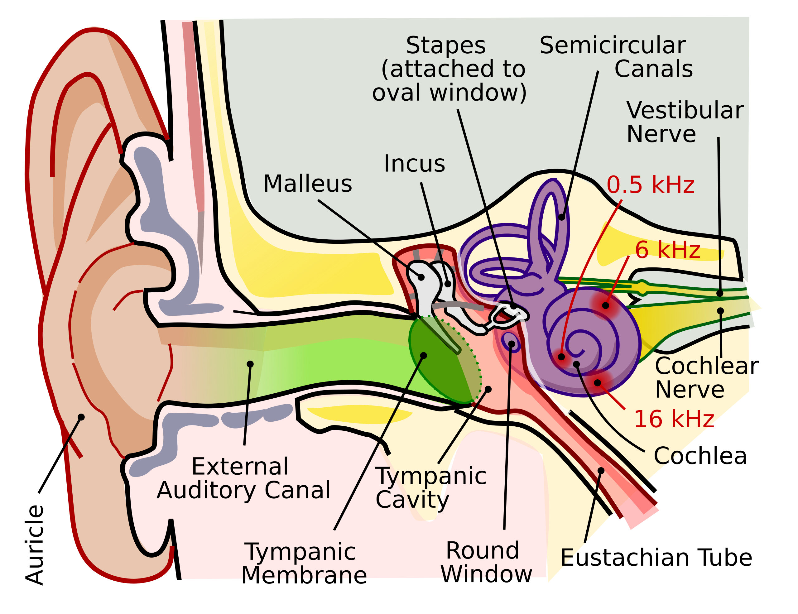
English: The human ear and frequency mapping in the cochlea. The three ossicles incus, malleus, and stapes transmit airborne vibration from the tympanic membrane to the oval window at the base of the cochlea. Because of the mechanical properties of the basilar membrane within the snail-shaped cochlea, high frequencies will produce a vibration peak near the oval window, whereas low frequencies will stimulate receptors near the apex of the cochlea (locations for three frequencies indicated schematically). Information from the cochlear receptor cells is transmitted to the cochlear nuclei via the 8th cranial nerve, and on through the midbrain to the cortex. |
| תאריך יצירה | |
| מקור | נוצר על־ידי מעלה היצירה (טקסט מקורי: “Own work by uploader, derived from File:Anatomy_of_the_Human_Ear.svg”) |
| יוצר | Inductiveload |
| אישורים והיתרים (שימוש חוזר בקובץ זה) |
הקובץ הזה מתפרסם לפי תנאי רישיון קריאייטיב קומונז ייחוס-שיתוף זהה 2.5 כללי.
|
| גרסאות אחרות |
[]
|
| SVGהתפתחות | Inkscape עם נוצרה ה גרפיקה וקטורית This file is translated using SVG switch elements: all translations are stored in the same file. |


היסטוריית הקובץ
ניתן ללחוץ על תאריך/שעה כדי לראות את הקובץ כפי שנראה באותו זמן.
| תאריך/שעה | תמונה ממוזערת | ממדים | משתמש | הערה | |
|---|---|---|---|---|---|
| נוכחית | 00:29, 17 בספטמבר 2018 | | 519 × 674 (33 ק"ב) | JoKalliauer | added systemLanguage="eo" |
| 20:21, 16 בספטמבר 2018 |  | 519 × 674 (32 ק"ב) | JoKalliauer | added systemLanguage="de" | |
| 08:33, 11 בספטמבר 2018 |  | 519 × 674 (87 ק"ב) | Jmarchn | Bigger (proportional real size) and full redraw (more realistic) of the auricle. Ossicles in white colour. Eardrum with contour. Added 3 labels. Add fundus to the bone and subcutaneous tissues, add superior auricular muscle, add transparency to middle ear, add separation between middle and inner ear, add division to internal auditory canal. | |
| 16:40, 29 באפריל 2009 |  | 600 × 800 (98 ק"ב) | Inductiveload | swap incus/malleus | |
| 18:10, 15 בפברואר 2009 |  | 600 × 800 (98 ק"ב) | Inductiveload | {{Information |Description={{en|1=The human ear and frequency mapping in the cochlea. The three ossicles incus, malleus, and stapes transmit airborne vibration from the tympanic membrane to the oval window at the base of the cochlea. Because of the mechan |
שימוש בקובץ
הדף הבא משתמש בקובץ הזה:
שימוש גלובלי בקובץ
אתרי הוויקי השונים הבאים משתמשים בקובץ זה:
- שימוש באתר en.wikipedia.org
- שימוש באתר en.wikibooks.org
- שימוש באתר eo.wikipedia.org
- שימוש באתר lt.wikipedia.org
- שימוש באתר www.wikidata.org